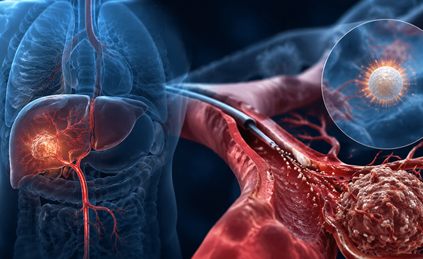
Radioactive microsphere embolisation, also known as selective internal radiation therapy, involves the precise injection of microspheres loaded with radionuclides (such as Yttrium-90) into the feeding arteries of liver tumours via microcatheters. The microspheres become permanently lodged in the terminal vascular bed of the tumour, continuously emitting high-energy beta rays to destroy tumour cells, whilst simultaneously producing a microvascular embolisation effect. As beta rays have an extremely short range within tissue, the radiation dose to normal liver parenchyma is controllable, thereby achieving internal radiation ablation of the tumour region.
High-dose local internal irradiation: The radiation dose in the target area is extremely high, whilst surrounding normal tissue is protected due to rapid radiation attenuation.
Synergistic effect of embolisation and radiotherapy: Blocking blood flow enhances the biological effects of radiation, making this approach particularly suitable for tumours with a rich blood supply.
Individualised dose planning: Radiation doses are precisely calculated preoperatively through simulated perfusion and radionuclide scans, ensuring treatment safety.
Primarily indicated for primary liver cancer and liver metastases that cannot be surgically resected, particularly in patients with portal vein tumour thrombi, poor hepatic reserve, or those unresponsive to chemotherapy. This approach can significantly prolong survival and improve quality of life.