Nasopharyngeal cancer shows marked geographic distribution, with high incidence in South China and Southeast Asia. EB virus infection is the main cause. Radiotherapy is the most important curative treatment, with overall survival exceeding 80%, but local recurrence and distant metastasis still require vigilance.
· EB virus infection
· South Chinese genetic background and specific HLA genotypes
· Salted fish and nitrite-containing foods
· Smoking and formaldehyde exposure
· Familial clustering
EB virus latent membrane proteins activate the NF-κB pathway. Against a background of genetic susceptibility, nitrites and other factors synergistically damage DNA, leading to malignant transformation of the nasopharyngeal epithelium.
Painless cervical lymph node enlargement is the most common presentation. Blood-stained postnasal discharge, unilateral ear fullness, and hearing loss are typical early warning signs. Advanced disease may cause nasal obstruction, headache, diplopia, and facial numbness.
· Surgery: Small localized recurrent lesions may be resected endoscopically through the nose; residual or recurrent cervical lymph nodes may be treated with neck dissection.
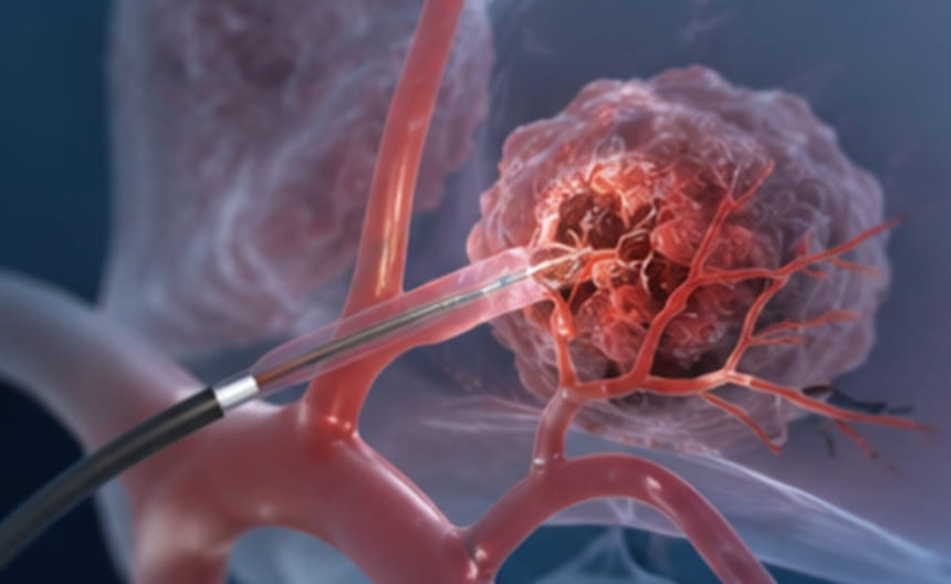
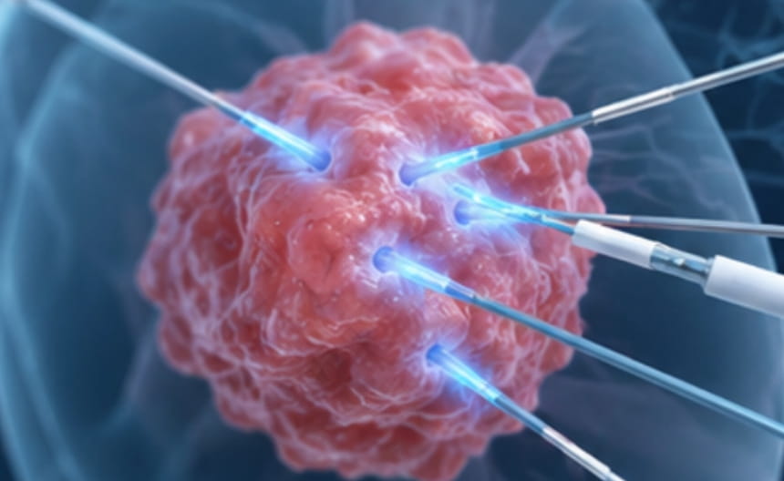
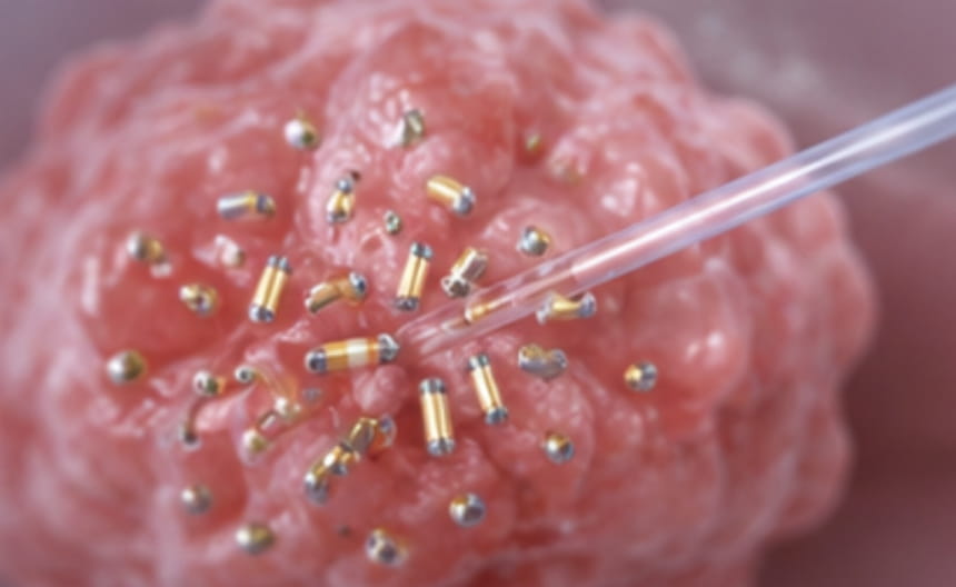
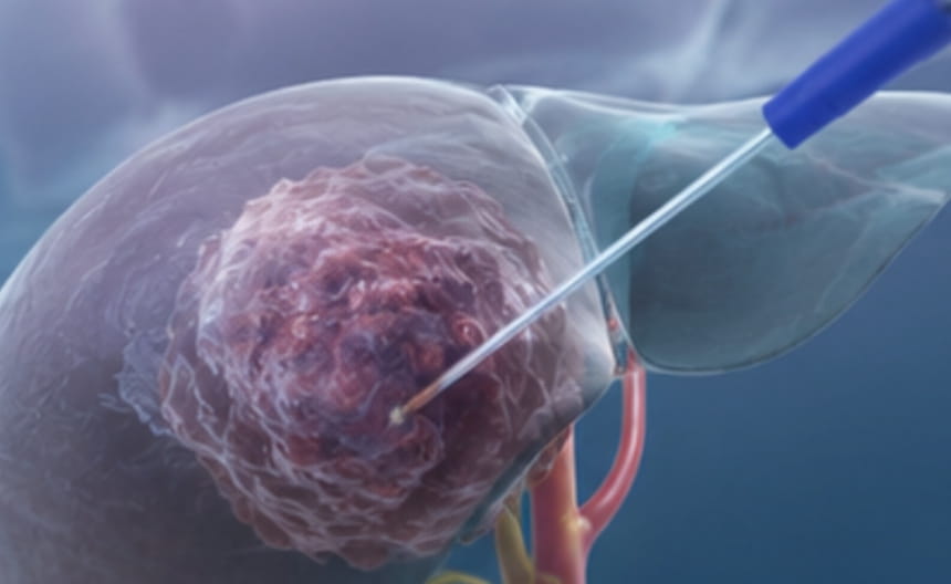
· Minimally invasive treatment: Local recurrent nasopharyngeal lesions may be treated with iodine-125 seed implantation for brachytherapy; image-guided radiofrequency ablation and microwave ablation may be used for isolated recurrent lesions.
· Chemoradiotherapy: Intensity-modulated radiotherapy combined with concurrent chemotherapy is the cornerstone of curative treatment; induction and adjuvant chemotherapy are used for locally advanced disease.
· Targeted and immunotherapy: Immune checkpoint inhibitors have shown good efficacy in recurrent or metastatic nasopharyngeal cancer.
· Others: EBV DNA is used to monitor treatment response and follow-up; nutritional support and protection of oral and hearing functions are integrated throughout treatment.
Diagnosis is confirmed by nasopharyngoscopy with biopsy. Plasma EBV DNA quantification is a sensitive marker for screening and monitoring. Head and neck MRI is the best method for local staging. PET-CT is used to evaluate distant metastasis.