
UNI-ASIA Logo WATA Logo

Minimally Invasive Care for Complex Vascular Malformations: Type-Specific Treatment by Liao Zhengyin’s MDT Team

  • 4 categories

    High-risk vascular malformationsComprehensive coverage

  • 24h

    High-risk shuntsTime to elimination

  • 48h

    MDT-signedDiagnosis and treatment report

SELF CHECK

Are you facing these moments of despair?

If any of these sound familiar, please read on

  1. You have visited many hospitals, only to hear "It is too complex to treat" or "Surgical removal is the only option".

  2. You have undergone interventional treatment or sclerotherapy, but your condition keeps worsening and recurring, with the vascular malformation returning aggressively.

  3. A facial malformation affects your appearance, leaving you afraid to go out or socialize, while constantly worrying about severe bleeding.

  4. Your doctor says the lesion must be removed extensively along with surrounding healthy tissue—an outcome you and your family cannot accept.

The "surgical no-go zones" others dare not touch Are precisely where we focus our efforts

WARNING

They may look alikeThe wrong treatment is like taking a scalpel to a bomb

Vascular malformations and ordinary hemangiomas are fundamentally different conditions

Ordinary hemangioma

Like a small, enclosed pouch of blood; most can subside with simple treatment

Complex vascular malformation

Like a network of misconnected water pipes; misguided treatment makes it more dangerous

Treating blindly only increases the danger

  1. Blocking the wrong site

  2. Blood flow balancecollapses instantly

  3. Malformed vesselsrebound aggressively

  • Mild

    Accelerated facial collapse and disfigurement, causing irreversible damage to appearance

  • Severe

    Massive internal bleeding that can become life-threatening at any moment

  • Critical

    The heart is pushed toward failure, affecting multiple organs

Blocking vessels blindly without understanding blood flow does not defuse the bomb—it sets it off

WHY US

The same condition, dramatically different outcomes

Map blood flow before treatment—a tiny difference can mean life or death

Conventional intervention Liao Zhengyin MDT team

When treating small, superficial malformations, healthy vessels may be embolized by mistake, causing disfigurement

Treat cranial and pelvic AVMs with superselective embolization, preserving facial appearance and organ function

Blind embolization of arteriovenous fistulas can cause fatal pulmonary embolism

Precisely occlude high-flow AV fistulas to reverse the progression of heart failure

Unable to manage diffuse venous malformations, with recurrence despite repeated injections

Combine percutaneous sclerotherapy under dual guidance with minimally invasive debulking to restore appearance

Failure to distinguish KTS from Parkes Weber syndrome may lead to unnecessary amputation

Use MDT differential diagnosis to identify the condition, block abnormal return flow and preserve limb function

TREATMENT

Four high-risk shunt types, targeted treatment for each

A "shunt" is an abnormal passage in the vascular systemOne strategy per type · Core strategies for elimination within 24 hours

Life-threatening high-flow shunts

Risk of death
Mechanism
Arterial blood flows directly through the malformation nidus into veins → cardiac volume overload → progressive heart failure
Conditions
Cranial AVM · Pelvic AVM · High-flow AV fistulas
Strategy
Destroy the nidus (ablation with absolute ethanol) · Cut off supply (embolization of feeding arteries through multiple routes) · Isolate the fistula (exclude its opening with a covered stent)
Expected results
Significantly reduced cardiac workload and restored organ perfusion

Extensive low-flow shunts

Disfigurement risk
Mechanism
Persistent blood leakage and fluid accumulation in a diffuse abnormal vascular bed → progressive local swelling and disfigurement
Conditions
Diffuse venous malformations of the face and neck · Blue rubber bleb nevus · Giant cystic lymphatic malformations
Strategy
Dual-guided targeting (precise puncture under combined ultrasound/MRI guidance) + Sclerotic closure (intralesional sclerotherapy combined with minimally invasive debulking)
Expected results
Bleeding stops completely and normal appearance is restored

Mixed-type shunts

Multisystem involvement
Mechanism
Simultaneous involvement of arterial, venous and lymphatic systems → progressive limb overgrowth and deformity
Conditions
CVM · CLVM (with soft tissue/bone overgrowth)
Strategy
Layered treatment: ① Embolize the arterial supply ② Sclerose venous lakes ③ Suppress lymphatic microcysts
Expected results
Enlarged limbs become noticeably slimmer and free movement is restored

Syndromic high-risk shunts

Often missed or misdiagnosed
Mechanism
Genetic mutations combined with hemodynamic disturbances → progressive multisystem deterioration
Conditions
KTS · Parkes Weber · PTEN hamartoma · CLOVES
Strategy
Treat the cause and its effects: Gene-targeted drugs to control the underlying cause + hemodynamic correction + sequential MDT management
Expected results
Preserve limbs and life, avoiding amputation and sudden death

ADVANTAGES

Liao Zhengyin MDT team: four core strengths

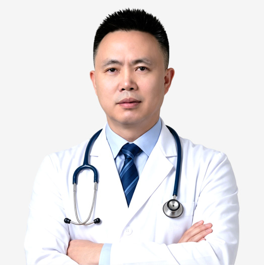
Liao Zhengyin

Liao Zhengyin Professor · PhD

A leading figure in multidisciplinary care for complex tumors in China

Former Director of the Cancer Center at West China Hospital · First-tier specialist

“ With complex vascular malformations, the issue is not "untreatable", but "not yet clearly understood"—once blood flow is fully understood, a no-go zone becomes a path forward. ”
  1. Map the vessels before treatment

    Trace blood flow from source to destination before the procedure

  2. Superselective embolization targets only the lesion

    Spare healthy vessels, preserving facial appearance and organs

  3. One plan developed by the MDT

    Specialists consult across disciplines to eliminate diagnostic errors

  4. Specialists in "no-go zone" cases

    Cases others hesitate to accept are routine here
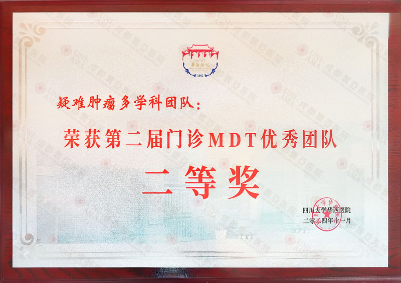
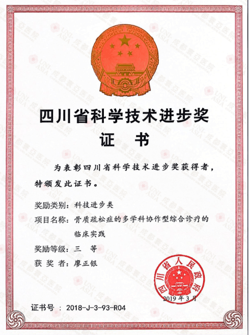
Outstanding MDT Team for Complex Tumors
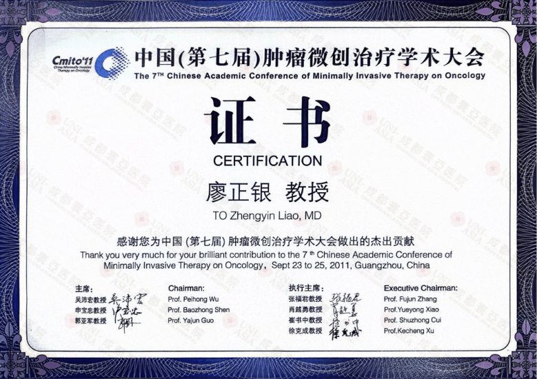
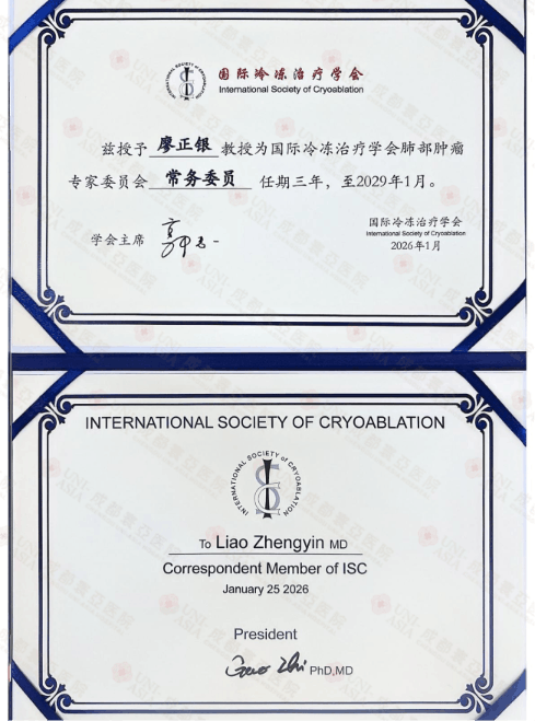
Chinese Minimally Invasive Association Certificate
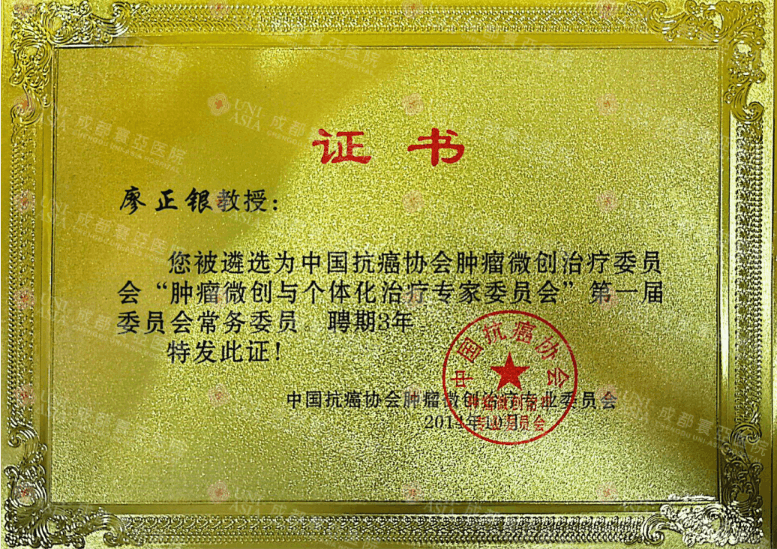
Member of the China Anti-Cancer Association
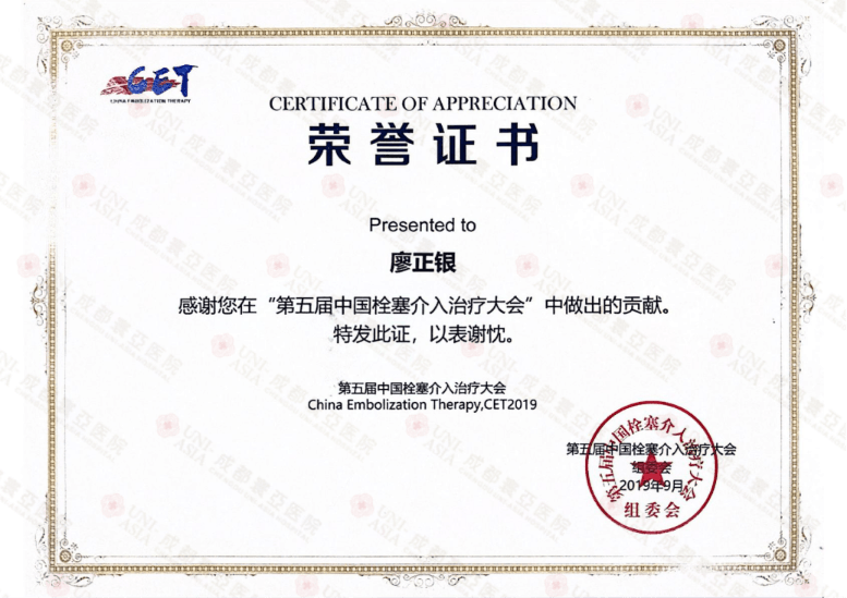
China Embolization Therapy Conference Certificate
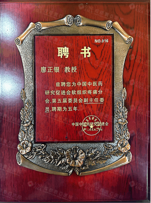
Deputy Director, Chinese Medical Research Association
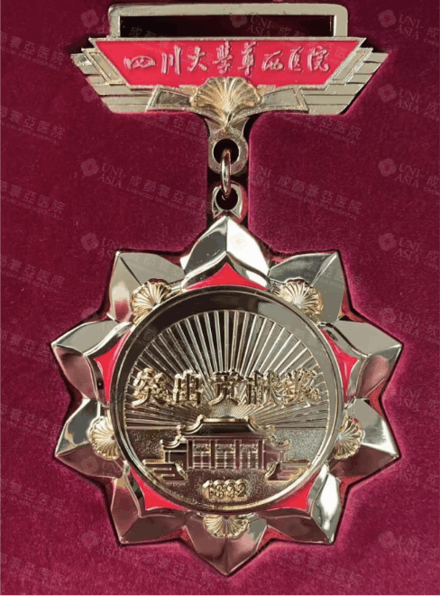
West China Hospital Outstanding Contribution Award

CASES

They were once told "nothing could be done"

These cases come from actual clinical recordsFor each patient told "nothing could be done", the story continued here

Arteriovenous malformation of the lip (Case 1)

57-year-old man / Left upper lip AVM (Yakes type IV)

Case 1 before treatment: pulsatile mass with blood crusting on the left upper lip
Before treatment: Pulsatile mass with blood crusting on the left upper lip
Case 1 at 1-year follow-up: swelling and pulsation largely resolved
1-year follow-up: Swelling and pulsation largely resolved
Background:
A pulsatile mass on the left upper lip had been present for years, with recurrent bleeding. Several hospitals recommended surgical excision, noting the risks of lip tissue loss and impaired appearance and function.
Treatment:
Interventional sclerotherapy and ablation with absolute ethanol (superselective embolization of a Yakes type IV AVM)
Outcome:
At 1-year follow-up, swelling and pulsation had resolved, with the lip contour preserved.

Arteriovenous malformation of the lip (Case 2)

38-year-old woman / Left lower lip AVM

Case 2 before treatment: marked thickening and a pulsating sensation in the left lower lip
Before treatment: Marked thickening and a pulsating sensation in the left lower lip
Case 2 at 1-year follow-up: lip shape largely restored
1-year follow-up: Lip shape largely restored
Background:
Progressive thickening of the left lower lip was accompanied by local pulsation and recurrent oral bleeding over several years. Another hospital recommended en bloc excision with immediate reconstructive surgery; the patient and her family declined this approach.
Treatment:
Percutaneous direct puncture of the abnormal vascular network + targeted embolization with absolute ethanol
Outcome:
Two months after treatment, abnormal local pulsation had resolved, with lip shape preserved and normal eating and speech.

Complex craniofacial arteriovenous malformation

Complex craniofacial AVM / Unsuccessful attempts to obtain treatment at multiple centers

Complex craniofacial AVM on pretreatment 3D vascular reconstruction: extensive abnormal vascular nidus throughout the craniofacial region
Pretreatment 3D vascular reconstruction: Extensive craniofacial vascular nidus
Complex craniofacial AVM on post-treatment DSA: occlusion of the abnormal vascular nidus
Post-treatment DSA: Abnormal vascular nidus occluded
Background:
The patient sought care at several medical centers for a complex craniofacial AVM. Each considered definitive treatment difficult because of the deep location and extensive involvement. Daily activities were substantially limited, and the platelet count was low.
Treatment:
Staged treatment with repeated transarterial embolization + direct-puncture ablation with absolute ethanol
Outcome:
Angiography showed occlusion of the main abnormal vascular nidus, with marked symptom improvement and no further recurrent bleeding.

Arteriovenous malformation of the forehead

Forehead AVM / Recurrence after surgery

Forehead AVM before treatment: purplish-black lesion with a high risk of ulceration
Before treatment: Purplish-black forehead AVM with a high risk of ulceration
Forehead AVM after interventional treatment: smaller lesion and resolved ulceration risk
After interventional treatment: Smaller lesion and resolved ulceration risk
Background:
A female patient with a forehead AVM had undergone surgical excision with poor results and residual scarring. Follow-up examination showed that the abnormal vascular network remained active, with a risk of ulceration and massive bleeding.
Treatment:
Superselective transarterial embolization + direct-puncture sclerotherapy (performed carefully to avoid feeding branches of the ophthalmic artery)
Outcome:
The bleeding risk resolved, the lesion shrank substantially, and both scarring and appearance improved.

These cases are drawn from actual clinical records of Professor Liao Zhengyin’s team and are provided for medical education and general health information only.

PROCESS

Consult online in three steps before visiting the hospital

Whether you are in China or abroadGet expert medical advice first, then decide whether to travel

A doctor writing medical notes
  1. Free consultation

    Submit your medical information online. Our specialist team completes an assessment within 30 minutes to tell you whether minimally invasive treatment is suitable for you.

  2. Upload your scans

    Submit original CT, MRI or other imaging online. Scans from overseas are also accepted for joint review by our multidisciplinary MDT team.

  3. Receive a signed report within 48 hours

    Treatment plan · Risk management plan · Cost estimate, issued and signed by Professor Liao Zhengyin’s team.

Review the report before deciding to visit—patients in China and abroad can avoid an unnecessary trip

FAQ

Before you visit, understand these three things

Other hospitals said my condition cannot be treated. Can you really treat it?

Complex cases that other hospitals hesitate to accept are part of our everyday practice: cranial and pelvic AVMs, high-flow AV fistulas, KTS and related conditions are routinely treated here. Start with a free online consultation. After assessment, our MDT team will explain whether treatment is possible and how it would be carried out. Confirm suitability before traveling, so you avoid an unnecessary trip.

Could minimally invasive treatment cause disfigurement or damage my organs?

Quite the opposite. The principle of superselective embolization is to "target only abnormal vessels while sparing healthy ones". Preserving facial appearance and organ function is the main distinction between our approach and conventional intervention.

I live in another city or overseas. How can a specialist review my scans first?

Simply submit your original CT, MRI or other imaging online; scans from overseas are also accepted. Within 48 hours, you will receive a report signed by Professor Liao Zhengyin’s team covering your treatment plan, risk management plan and cost estimate. Review it before deciding whether to visit.

Weekly MDT consultation slots are limited · Submit early for an earlier appointment

Do not let "untreatable" be the final answer

Start with a free consultation and receive a signed report within 48 hoursWhether treatment is possible, how it works and the estimated cost—all explained clearly

Free medical record review · Book our expert team

Many patients once told "nothing can be done" have found a turning point here

Consultation

Name
Age
Phone Number
Email
Your Question

Submitted Successfully

We have received your consultation. Thank you for your trust. A specialist will contact you within 72 hours, so please keep an eye on your phone calls and email.

Book Appt. WhatsApp
Call Us